
Table of Contents

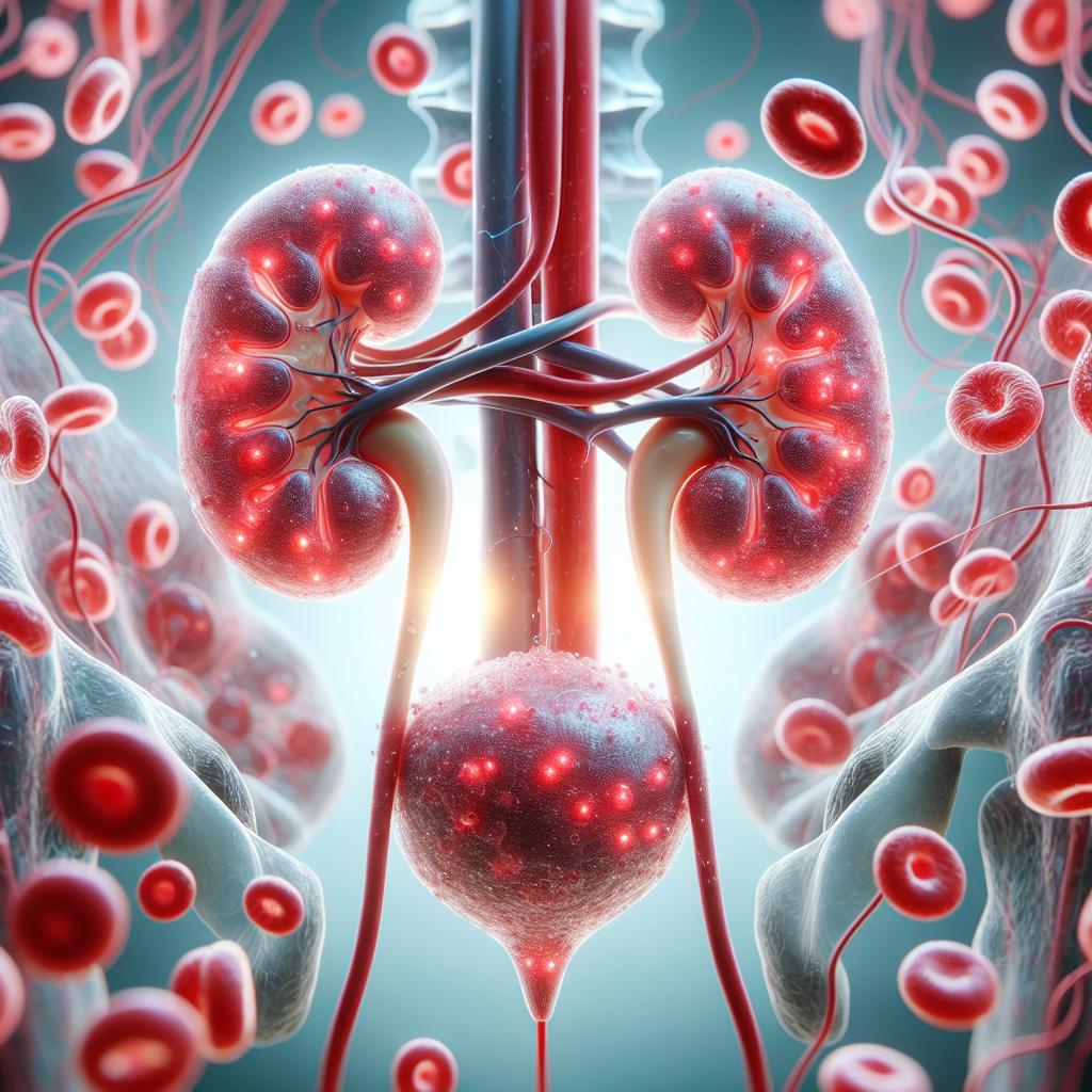
Definition and Overview of Microscopic Hematuria
Microscopic hematuria is defined as the presence of red blood cells (RBCs) in the urine that is not visible to the naked eye but can be detected through microscopic examination. While hematuria can be classified as either gross or microscopic, the latter is often discovered incidentally during routine urinalysis, raising concerns for potential underlying conditions. The prevalence of microscopic hematuria varies, with studies suggesting rates ranging from 1% to 5% in the general population, and higher in specific groups such as those with urological symptoms or risk factors (Mombo-Ngoma et al., 2024).
Microscopic hematuria may be transient or persistent. Its significance largely depends on the associated clinical context, as it can indicate benign conditions or serious underlying pathologies, such as urinary tract infections, kidney stones, or malignancies. Therefore, understanding the potential causes, conducting appropriate diagnostic evaluations, and implementing effective management strategies are crucial in addressing this condition.
Common Causes of Microscopic Hematuria in Adults
The causes of microscopic hematuria can be broadly categorized into renal and extrarenal origins.
Renal Causes
- Glomerular Diseases: These include conditions such as IgA nephropathy, minimal change disease, and glomerulonephritis. They often present with proteinuria or edema alongside hematuria.
- Urological Conditions: Renal stones, tumors, and cysts can contribute to blood in the urine. Urothelial carcinoma, particularly in older adults, has a significant association with hematuria.
- Infections: Urinary tract infections (UTIs) can cause irritation and bleeding of the urinary tract lining, leading to hematuria.
- Vascular Issues: Conditions like renal artery stenosis or embolism may result in ischemia and subsequent hematuria.
Extrarenal Causes
- Systemic Diseases: Conditions like hypertension and diabetes can lead to secondary kidney damage, manifesting as hematuria.
- Medications: Certain medications, including anticoagulants and nonsteroidal anti-inflammatory drugs (NSAIDs), can increase the risk of bleeding.
- Trauma: Any direct injury to the kidneys or urinary tract may result in hematuria.
Malignancies
The presence of microscopic hematuria can be particularly concerning in the context of malignancies. Bladder and renal cancers are significant considerations, especially in patients over 40 years of age. The risk of cancer increases with the persistence of hematuria, particularly if associated with other urinary symptoms (Thacharodi et al., 2024).
Diagnostic Approach to Microscopic Hematuria
The diagnostic evaluation of microscopic hematuria typically follows a systematic approach to identify the underlying cause.
Initial Assessment
- History and Physical Examination: A comprehensive medical history and physical examination are crucial. Important factors include age, gender, previous urinary symptoms, and family history of renal diseases.
- Urinalysis: A urinalysis is conducted to confirm hematuria and assess for protein, glucose, leukocytes, and nitrites. Dipstick tests may provide preliminary findings, but microscopic confirmation is necessary.
Further Investigations
- Imaging Studies: After initial evaluation, imaging studies such as renal ultrasound or computed tomography (CT) scans are recommended. These tests help visualize structural abnormalities within the kidneys and urinary tract.
- Cystoscopy: In cases where malignancy is suspected, especially in older adults or those with risk factors, cystoscopy allows for direct visualization of the bladder and urethra, enabling biopsy if necessary.
- Laboratory Tests: Blood tests may be ordered to evaluate renal function, electrolyte levels, and assess for potential systemic causes such as coagulopathy or glomerular diseases.
Risk Stratification
Patients are categorized based on the findings from history, physical examination, urinalysis, and imaging studies. Those with persistent or symptomatic hematuria warrant further investigation to rule out serious conditions, including malignancies.

Treatment Options for Microscopic Hematuria
The treatment of microscopic hematuria is contingent upon the underlying cause identified through diagnostic evaluation.
Management of Underlying Conditions
- Infections: If a UTI is diagnosed, appropriate antibiotic therapy is initiated. Follow-up urinalysis is essential to ensure resolution of hematuria.
- Nephrolithiasis: Patients with kidney stones may require hydration, analgesics, and in some cases, surgical interventions such as lithotripsy or ureteroscopy to remove stones.
- Glomerular Diseases: The management of conditions like glomerulonephritis may involve corticosteroids and immunosuppressants depending on the severity and specific diagnosis.
- Malignancies: Referral to urology for further management, including possible surgical intervention or chemotherapy, is necessary for patients with identified tumors.
Supportive Care
In cases without serious underlying pathology, management may involve lifestyle modifications, including increased fluid intake, avoiding nephrotoxic medications, and regular monitoring.
Importance of Follow-up in Microscopic Hematuria Cases
Regular follow-up for patients with microscopic hematuria is vital for several reasons:
- Monitoring for Recurrence: Patients may experience recurrence of hematuria, necessitating ongoing surveillance to detect potential underlying conditions early.
- Adjustment of Treatment: Changes in symptoms or urinary findings may require adjustments in treatment or further investigations.
- Patient Education: Providing education regarding the importance of reporting new symptoms, maintaining follow-up appointments, and adhering to management plans is crucial for patient outcomes.
FAQs
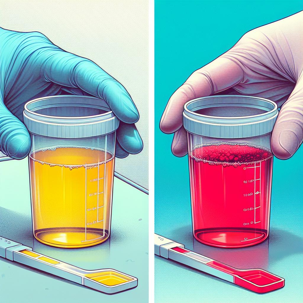
What is the difference between microscopic and gross hematuria?
Microscopic hematuria cannot be seen by the naked eye and is detected through urine tests, while gross hematuria is visible, with the urine appearing red or brown.
Is microscopic hematuria serious?
Microscopic hematuria can indicate serious underlying conditions, including infections, stones, or malignancies. Therefore, further evaluation is important to determine the cause.
How is microscopic hematuria diagnosed?
It is diagnosed through urinalysis, which confirms the presence of red blood cells in the urine, followed by further diagnostic testing to identify any underlying conditions.
What are common treatments for microscopic hematuria?
Treatment depends on the underlying cause but may include antibiotics for infections, lifestyle changes for stones, or immunosuppressants for glomerular diseases.
When should I see a doctor for hematuria?
You should seek medical attention if you notice blood in your urine, experience pain during urination, or have other urinary symptoms.
References
- Mombo-Ngoma, G., et al. (2024). Asymptomatic Malaria Infection and Hidden Parasitic Burden in Gabonese Schoolchildren: Unveiling Silent Co-Infections in Rural and Urban Settings. Tropical Medicine and Infectious Disease. https://doi.org/10.3390/tropicalmed10010011
- Thacharodi, A., et al. (2024). The burden of group A Streptococcus (GAS) infections: The challenge continues in the twenty-first century. iScience. https://doi.org/10.1016/j.isci.2024.111677


