
Table of Contents

Importance of Urine Dipstick in Routine Medical Checkups
The urine dipstick test is an invaluable tool in routine medical checkups, providing quick and efficient insights into various health conditions. This simple, cost-effective test can screen for a variety of health issues, including urinary tract infections (UTIs), kidney disease, and metabolic disorders. Its significance lies in its ability to deliver rapid results, allowing healthcare professionals to make informed decisions about further diagnostic steps or treatments.
Urine dipstick testing typically involves the use of colorimetric strips that change color in response to various substances in the urine. These strips can detect parameters such as glucose, protein, ketones, bilirubin, and hemoglobin, among others. For instance, the presence of glucose can indicate diabetes mellitus, while proteinuria may suggest kidney damage or disease. Regular urine dipstick testing is particularly important for at-risk populations, such as individuals with diabetes, hypertension, or a family history of kidney disease.
Research indicates that early detection of abnormalities through urine dipstick tests can lead to prompt intervention, reducing the risk of severe complications (Huang et al., 2025). For example, in patients with diabetes, monitoring urine glucose levels can help manage blood sugar levels more effectively, thereby preventing the long-term complications associated with uncontrolled diabetes.
How to Properly Use a Urine Dipstick for Accurate Results
To ensure accurate results when performing a urine dipstick test, proper technique and timing are essential. Here are the steps involved in correctly using a urine dipstick:
-
Collection of Urine Sample: The urine sample should be collected midstream to minimize contamination. Patients are typically advised to clean the genital area before collection to ensure the sample’s integrity.
-
Using the Dipstick: After collecting the urine, the dipstick is immersed into the sample for a specified amount of time (usually a few seconds). It is crucial not to leave the dipstick in the urine for too long as this can lead to inaccurate results due to diffusion of reagents.
-
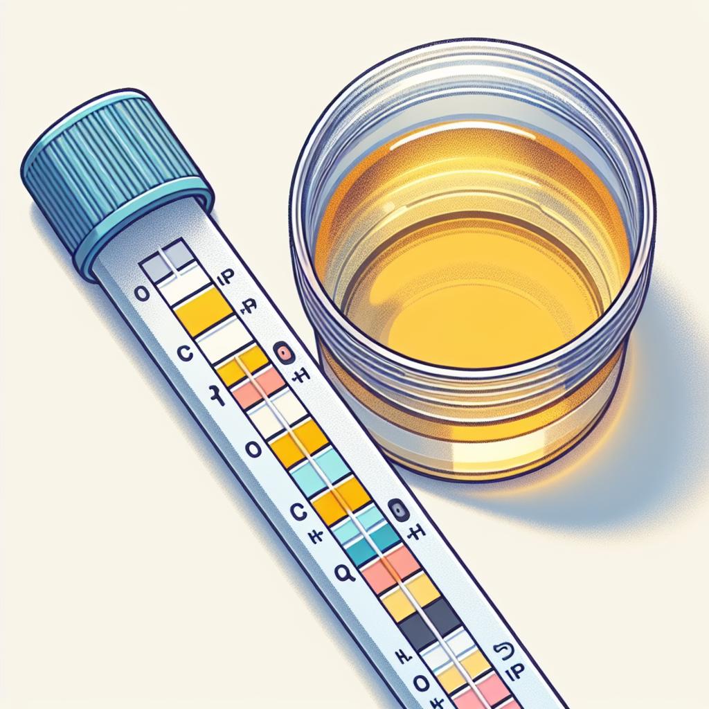
Reading the Results: After removing the dipstick, it should be held horizontally to prevent the reagents from running into adjacent pads. The results should be read at the time specified by the manufacturer, as different analytes react at different rates.
-
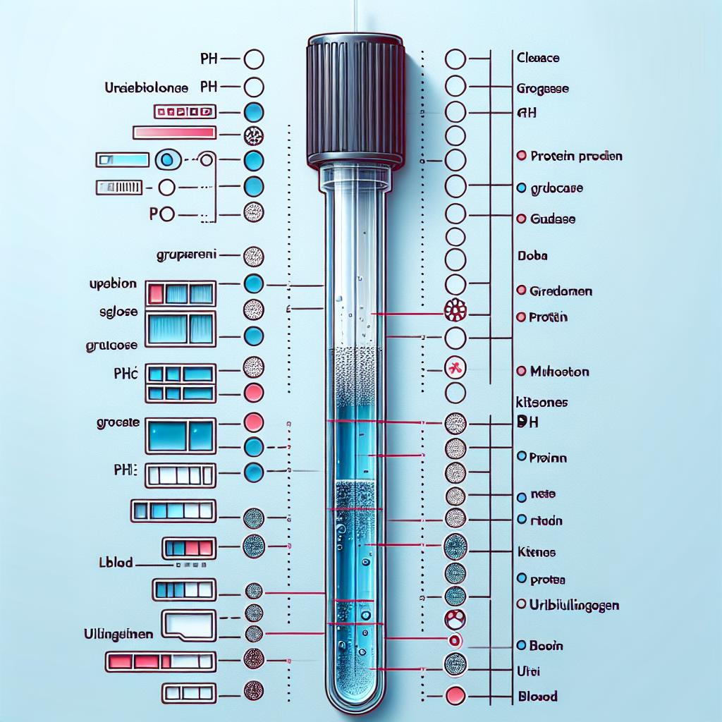
Comparison to Color Chart: Each pad on the dipstick corresponds to a specific analyte and has an associated color chart. The color of each pad is compared to the chart to determine the concentration of the substance in the urine.
-
Documentation and Follow-Up: All results should be documented, and any abnormal findings should prompt further investigation or follow-up with healthcare professionals.
In clinical settings, adherence to these guidelines is paramount. Variability in technique, such as improper sample collection or timing, can lead to erroneous interpretations which may mislead diagnosis or treatment plans (Mugusi et al., 2025).
Common Conditions Detected by Urine Dipstick Testing
Urine dipstick tests are effective in screening for a variety of conditions. The following are some of the common conditions detected through this method:
- Diabetes Mellitus: The presence of glucose in the urine indicates uncontrolled diabetes or renal glycosuria, prompting further blood glucose testing.
- Urinary Tract Infections (UTIs): Nitrites and leukocyte esterase are both indicators of bacterial infections in the urinary tract. A positive result necessitates additional cultures and sensitivity testing.
- Kidney Disease: Proteinuria can indicate kidney damage or disease, such as nephrotic syndrome or glomerulonephritis. Monitoring protein levels can help assess the effectiveness of treatment in patients with known kidney conditions (Pinto et al., 2024).
- Liver Dysfunction: Bilirubin in the urine suggests liver disease or hemolysis, necessitating further liver function tests.
- Hematuria: The presence of blood can be indicative of various conditions, including infections, kidney stones, or malignancies in the urinary tract.
Table 1 summarizes the common conditions and their corresponding urine dipstick indicators:
| Condition | Urine Dipstick Indicator |
|---|---|
| Diabetes Mellitus | Glucose |
| Urinary Tract Infection | Nitrites, Leukocyte Esterase |
| Kidney Disease | Protein |
| Liver Dysfunction | Bilirubin |
| Hematuria | Blood |
Advantages of Urine Dipstick Over Other Diagnostic Methods
Urine dipstick testing offers numerous advantages compared to other diagnostic methods, making it an essential tool in healthcare:
-
Cost-Effectiveness: Urine dipsticks are inexpensive compared to more complex diagnostic tests, making them accessible for routine screenings in various healthcare settings.
-
Rapid Results: The test provides immediate results, allowing for quick clinical decision-making and timely interventions.
-
Simplicity: The procedure is straightforward and can be performed by healthcare professionals with minimal training. This ease of use allows for widespread application in outpatient settings.
-
Non-Invasive: Collecting a urine sample is less invasive than blood draws or biopsies, reducing patient discomfort and anxiety.
-
Comprehensive Screening: A single urine dipstick test can screen for multiple conditions simultaneously, enhancing the efficiency of patient evaluations.
-
Monitoring Disease Progression: For patients with chronic conditions, regular urine dipstick testing can help monitor disease progression or response to treatment. For instance, patients with diabetes can track their renal function and glycemic control over time (Gadzhiev et al., 2025).
Interpreting Urine Dipstick Results: A Guide for Patients
Understanding urine dipstick results can empower patients to take an active role in managing their health. Here’s a guide to interpreting common urine dipstick results:
- Negative Results: A negative test for glucose, protein, and blood indicates normal renal and metabolic function.
- Positive Glucose: May indicate diabetes or renal glycosuria. Patients should consult their healthcare provider for further evaluation.
- Positive Protein: Indicates potential kidney damage. Further tests, such as a 24-hour urine collection, may be necessary for accurate assessment.
- Positive Nitrites and Leukocyte Esterase: Suggest an infection. Patients may require antibiotic therapy based on culture results.
- Positive Bilirubin: Implies liver dysfunction or hemolytic anemia. Liver function tests will be needed for a definitive diagnosis.
- Positive Blood: May indicate infections, stones, or malignancies. Additional imaging or cystoscopy could be required to identify the underlying cause.
Patients should be encouraged to discuss any abnormalities with their healthcare provider for appropriate follow-up and management (Sadi et al., 2021).

FAQ Section
How often should I have a urine dipstick test?
The frequency depends on individual health conditions. Patients with diabetes or kidney disease should have regular screenings as advised by their healthcare provider.
Are there any risks associated with urine dipstick testing?
No, urine dipstick testing is non-invasive and carries no significant risks. However, it is crucial to ensure proper collection techniques to avoid contamination.
Can I perform a urine dipstick test at home?
Yes, home urine dipstick testing kits are available, but it’s essential to follow the instructions carefully and consult with a healthcare provider for interpretation.
What should I do if I receive abnormal results?
Contact your healthcare provider promptly for further evaluation and potential follow-up testing.
References
-
Huang, R., Jiang, M., Chen, J., Cao, Z., Wang, Z., Ma, Z., Lin, G. (2025). Flexible ureteroscopy combined with potassium sodium hydrogen citrate intervention improves the stone-free rate for 20–30 mm uric acid renal stones. BMC Urology, 25(1), 64. https://doi.org/10.1186/s12894-025-01710-0

-
Mugusi, S. F., Shayo, G. A., Sasi, P. G., Fundikira, L. S., Aris, E. A., Sudfeld, C. R., Mugusi, F. M. (2024). Kidney disease among adults on tenofovir-based second-line antiretroviral therapy in Dar es Salaam, Tanzania. South African Journal of HIV Medicine, 26(1), 1640. https://doi.org/10.4102/sajhivmed.v26i1.1640
-
Pinto, V. M., Cima, R., Di Maggio, R., Alga, M. L., Gigante, A., Longo, F., Pasanisi, A., Venturelli, D., Cassinerio, E., Origa, R., Zanconato, G., Forni, G. L. (2024). Thalassemias and Sickle Cell Diseases in Pregnancy: SITE Good Practice. Journal of Clinical Medicine, 14(3), 948. https://doi.org/10.3390/jcm14030948
-
Sadi, M. V., Saltzman, N., Feria, G., Gittes, R. F. (2021). Experimental observations on dissolution of uric acid calculi. Journal of Urology, 134(3), 575-579 17)47303-4
-
Gadzhiev, N. K., Obidnyak, V. M., Gorelov, D. S., Malkhasyan, V. A., Akopyan, G. N., Mazurenko, D. A., Kharchilava, R. R., Petrov, S. B., Martov, A. G. (2025). A retrospective study comparing super-mini percutaneous nephrolithotomy and flexible ureteroscopy for the treatment of 20–30 mm renal stones in obese patients. PeerJ, 8, e8532. https://doi.org/10.7717/peerj.8532
-
Mugusi, S. F., Shayo, G. A., Sasi, P. G., Fundikira, L. S., Aris, E. A., Sudfeld, C. R., Mugusi, F. M. (2024). Kidney disease among adults on tenofovir-based second-line antiretroviral therapy in Dar es Salaam, Tanzania. South African Journal of HIV Medicine, 26(1), 1640. https://doi.org/10.4102/sajhivmed.v26i1.1640


